Tous droits réservés © NeurOreille (loi sur la propriété intellectuelle 85-660 du 3 juillet 1985). Ce produit ne peut être copié ou utilisé dans un but lucratif.
 Français
 English
 Español
 Português
The structure and function of the organ of Corti vary depending on three factors: (1) the position along the cochlear spiral (base to apex); (2) the frequency specialisation of the species of interest and; (3) its development (described in specific pages).
Variations relating to cochlear position (base-apex)
In transverse sections
")
M. Lenoir  et apical (haut) d'une cochlée de cobaye")
R Pujol |
Basal (bottom) and apical (top) turns of a guinea pig cochlea. The main morphological differences between the base and the apex are that:
Scale bar: 20 µm |
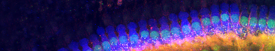
Surface views using scanning electron microscopy (SEM)

M Lenoir |
Organisation of hair cells in the apex of a rat cochlea |

M Lenoir |
Arrangement of hair cells at the base of the cochlea
Note the regular arrangement of the hair cells and their short stereocilia. Scale bar: 15 µm |
Outer hair cell stereocilia imprints in the tectorial membrane
The tallest OHC stereocilia are firmly implanted in the tectorial membrane. This is another characteristic 'base-apex' difference, as imprints cannot be seen in the apical part of the cochlea.

M Lenoir 
|
Inferior side of the tectorial membrane at the base of a rat cochlea The W-shaped imprints of the longest stereocilia of the three rows of OHCs are highly visible, particularly at higher magnification (below). Scale bar: 10µm |

R. Pujol |
Electron microscope section of an OHC slightly removed from its slot (red arrow) in the tectorial membrane. |
The embedding of OHC stereocilia in the tectorial membrane reinforces the coupling between the OHCs and the surrounding structures. It makes the cells’ active mechanisms and transduction much more efficient.
Interspecies variations relative to frequency specialisation

Cochlear structure is relatively stable in mammals, but depending upon the perceived frequency range, differences in the outer hair cells can be observed between species. Notably, the length of the OHCs and their stereocilia increases gradually from the base to the apex between species (see figure below). At each end of this frequency specialisation continuum, ultra-specialised structures can be seen for very low (mole) or very high (bat, dolphin) frequencies.
The "xylophone" shows the OHC length variation according to the coded frequency

Schematic drawing representing OHCs from different mammalian species and different cochlear turns. While OHC diameter keeps a constant value (7 µm), their length regularly varies according to frequency. In the human cochlea, a 25 µm basal OHC (C) is found at a place which codes for 20 kHz; conversely a 70 µm OHC (G) is found apically at the site coding for a very low frequency (< 100 Hz). A = shortest OHC in basal turn of a bat cochlea (at a place coding for 160 kHz), B = basal OHC from a cat cochlea (at a place coding for 40 kHz), D = OHC from second turn of a guinea pig cochlea (at a place coding for 5 kHz), E = OHC from start of third turn of a guinea pig cochlea (at a place coding for 2.5 kHz), F = OHC from end of third turn of a guinea pig cochlea (at a place coding for 150 Hz), H = apical OHC from a mole rat cochlea (at a place coding for 15 Hz).

M. Lenoir |
Organisation of hair cells at the extreme apex of a mole cochlea
The disorganisation of the extreme apex of the cochlea is more pronounced in animals adapted to hearing extremely low sounds, such as the mole. Here, only the row of IHCs is visible, as though the OHCs were of no use at these frequencies (< 10 Hz). |

M. Vater et M. Lenoir |
Organisation of hair cells at the extreme base of a bat cochlea The organ of Corti of a bat cochlea is hyperspecialised for very high frequencies: at the extreme base, IHCs are very widely spaced, whereas the three rows of OHCs are very regular. Could the proportion of 1 IHC to 6-9 OHCs be required to code frequencies greater than 100 kHz? Scale bar: 15 µm |
Developmental variations
See specific pages " Development of the cochlea" and " Outer hair cells synaptic variations"
Facebook Twitter Google+