IHCs: physiology
Authors: Régis Nouvian
Contributors: Rémy Pujol, Sam Irving
Inner hair cells code the frequency and intensity of acoustic stimuli into a neural signal, which is then transmitted to the brain via the auditory nerve.
Note: Figure legends are temporarily in French
Frequency coding: Phase locking
Frequency coding in the mammalian cochlea is linked to cochlear tonotopy. However, for sounds with low frequencies (under 3 kHz), which are encoded by the apex of the cochlea, frequency coding depends on a second mechanism: phase locking at the level of the IHC.
Acoustic stimulation causes the stereocilia to deflect, stretch-sensitive ion channels open, and this causes an influx of cations into the hair cell. The latter depolarises, causing voltage-sensitive calcium channels to open. The influx of calcium promotes the release of glutamate into the synaptic cleft, effectively activating the auditory nerve fibres and resulting in action potentials.
Acoustic stimulation at 500 Hz
- The receptor potential manifests as an alternating depolarisation-repolarisation of the cell, which follows each phase of the acoustic stimulus frequency. The alternating current (AC) of the receptor potential is dominant. The direct current (DC) is negligible.
- Each time a hair cell depolarises, in phase with the stimulation frequency, glutamate is released. This leads to the generation of action potentials at a preferential moment in the stimulation phase: the nerve fibres are therefore responding in phase with the acoustic stimulus.
Acoustic stimulation at 2000 Hz
- IHCs are not fast enough to alternate between depolarisation-repolarisation cycles in phase with the stimulation frequency. The alternating component of the receptor potential decreases whereas the direct component continues to increase.
- The increase in the stimulation frequency (above 3 kHz) causes a loss of synchrony between the action potentials and the phase of the stimulation frequency.
Acoustic stimulation at 5000 Hz
- The receptor potential corresponds only to the direct component (DC). IHCs code the envelope of the sound stimulus and not stimulation frequency.
- Action potentials are no longer synchronised with stimulation, but are essentially generated at the beginning of the stimulus.
Intensity coding
In response to acoustic stimulation of increasing intensity, neuronal activation (the number of action potentials) increases with sound intensity. This is known as intensity coding.
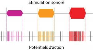
Spontaneous neural activity (in black) occurs without acoustic stimulation.
For three stimuli with increasing intensity (purple, orange and red), the evoked response (indicated by the dotted lines) is composed of an increasing number of action potentials.
Dynamic intensity coding
Auditory nerve fibres can be divided into three types:
- sensitive fibres (with low thresholds) and a high spontaneous firing rate (1)
- fibres with average thresholds and spontaneous firing rates (2)
- fibres with high thresholds and a low spontaeous firing rate (3).
Fibres with low thresholds and high spontaneous firing rates (1) have wide post-synaptic densities and are connected to small synaptic ribbons. On the contrary, high-threshold fibres with low spontaneous activity (3) have narrow post-synaptic densities and are connected to large synaptic ribbons.
The amplitude of the calcium influx (represented at the bottom of the presynaptic ribbon) is correlated with the spontaneous activity of the nerve fibres.

R Nouvian
Progressive recruitment of these three populations of nerve fibres can explain the extent of cochlear dynamics from perceptual threshold to the pain threshold (110 dB at 1000 Hz): in response to a weak stimulation, the low-threshold fibre (1, in red) is activated but becomes saturated very quickly. More intense stimulation causes the recruitment of fibres 2 and 3. Note that spontaneous activity occurs in absence of acoustic stimulation (black). Dotted lines indicate the beginning and the end of the acoustic stimuli, and isolate the evoked activity of the fibres.
Evoked activity of a fibre connected to the IHC
The post-stimulus time histogram (PSTH) shows evoked activity in response to acoustic stimulation. It is characterised by an increase in activation at the beginning of the stimulus (‘on response’), followed immediately by a decrease in evoked activity (‘adaptation’) until it reaches a constant discharge rate (‘plateau’).

The on response corresponds to the release of the neurotransmitter glutamate by the IHCs.
The adaptation can reflect: (i) the activation of the rapid IHC potassium conductance, (ii) the depletion of the fast-releasing synaptic vesicles, (iii) the desensitisation of post-synaptic glutamate receptors, and (iv) the refractory period of the action potential.
Stopping the acoustic stimulation is associated with a drastic decrease in the fibre’s discharge rate. This ‘off’ response could reflect the excessive glutamate recapture by the membrane transport proteins.