Audiometry
Authors: Jérôme Ruel, Nuno Trigueiros-Cunha, Jean-Luc Puel
Contributors: Sam Irving
Hearing thresholds of an individual may be assessed by both objective and subjective methods. Objective methods are based on recordings of the electrical activity at various stages along the auditory pathway, from the cochlea to the cortex, and do not need the active participation of the individual. On the other hand, subjective methods need the co-operation of the individual being tested, because they rely on the subject’s perception of and response to sound.
Objective methods
Auditory potentials reflect the electrical activity of the various neural pathways involved in the coding of sounds. Individual unitary potentials are recorded directly from a single cell or nerve fibre. Compound potentials, recorded at a distance from their origin, reflect the sum of the unit potentials.
The following are three examples of different types of potentials : note the temporal concordance beween the different recordings – at whichever level being measured, the first wave recorded with a latency of 1 ms reflects the auditory nerve action potential. Recording acoustic otoemissions is also an objective method, which specifically allows to check outer hair cells motile properties.
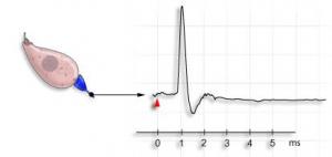
Unit action potentials (experimental technique not used in humans)
A micro-electrode (black arrow) records electrical activity from a single neurone in the auditory nerve. This auditory fibre responds to sound stimulation (red dot) by sending an action potential (AP) towards the brain. The latency between the sound stimulus (red arrow) and the the AP is about 1ms.
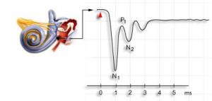
Recording of the compound cochlear nerve action potential at the Round Window.
This technique is used in human subjects, and is also known as electrocochleography.
A transtympanic electrode is placed on the promontory, near to the cochlear Round Window, and it records the compound action potential (CAP) of the auditory nerve, which is the synchronous activity of all the individual unit action potentials after sound stimulation (red point).
This potential is more complex than the single unit AP, but the first wave in this potential (N1) corresponds to the single unit AP in the preceding example. The amplitude of the CAP measured between N1-P1 is in the order of several dozen microvolts (µV). Successive responses are added and then averaged to eliminate background electrical noise.
Brainstem Auditory Evoked Potentials (BAEPs)
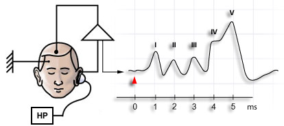
Surface electrodes placed on the skull allow recording of 5 main waves of electrical activity through the brainstem. Wave I, with a latency of 1 ms, reflects the auditory nerve potential ; waves II to V reflect processing centres in the auditory system.
These potentials are of very weak amplitude (< µV) and therefore averaging of many repeat stimulations (1000 to 2000) is required, in order to be able to differentiate them from background noise. After these early evoked potentials, the same skull electrodes can record later potentials from activity in the thalamocortex (middle latency and cortical auditory evoked responses).
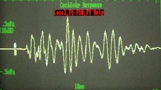
Oto-acoustic emissions (OAEs)
They reflect the activity of outer hair cells, which are the most sensitive cells of the organ of Corti.
OAEs are very useful for screening newborns, but are also widely used as as an objective test in subjects of any age.
Subjective methods (Audiograms)
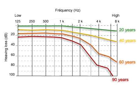
Tonal audiogram
Pure tone audiograms recorded in a normally-hearing subject over the course of their life. Each curve represents the average hearing loss as a function of age.
(see pathology / presbycusis)
Note: each one of these methods is further developed in specific pages