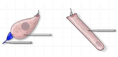
Cochlea
Authors: Jérôme Ruel, Nuno Trigueiros-Cunha, Jean-Luc Puel
Contributors: Patrick Minary, Sam Irving
To evaluate the cochlear function there are a number of objective methods. Some recordings, as unit potentials, are restricted to experimental approaches in animals. Other recordings, as electrocochleography, may be realized in human subjects.
Oto-acoustic emissions are developed in a specific page.
Cochlear potentials
There are two types of cochlear potentials : individual unit potentials recorded directly from a sensory cell or nerve cell, and compound potentials, recorded at a distance, reflecting the activity of several cells.
Single unit potentials
Les potentiels unitaires des cellules sensorielles sont aussi appelés potentiels de récepteurs.
Single unit potentials recorded from the sensory cells are also known as receptor potentials.
These potentials are recorded via a micro-electrode placed directly into the cell (intracellular recordings).Single unit potentials of the primary auditory neurones can be recorded either at the level of the dendrites (as shown), the cell bodies in the spiral ganglion, or at the level of the auditory nerve fibres.
Hair cell receptor potentials
The electrical response of the hair cells to an acoustic stimulus (shown at the bottom of the figure) is made up of two components : a continuous component (CC), duplicating the acoustic stimulus envelope, and an alternating component (AC), superimposed on the CC, corresponding to the pure sound frequency.
These continuous and alternating components depend on the frequency of sound stimulation. The amplitude of the CC grows with the frequency, whereas the CA is more important for low frequencies. The CA is more important in the outer hair cells than the inner hair cells, and the CC is more dominant in the inner hair cells (ref. Russell et al.).
Auditory nerve single unit potentials
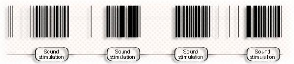
A sound stimulation (click) leads to an increase in the numbre of single unit potentials in the individual fibres of the auditory nerve. This increase in the rate of discharge, synchronised with the sound stimulation, is called an evoked response. The presence of action potentials during periods of silence is a reflection of the basal activity of the nerve fibre, also known as its spontaneous activity.

Electrocochleography (ECochG) is the name given to the recording of cochlear potentials.
Under local anaesthetic, a thin needle electrode is placed through the tympanic membrane onto the promontory, near the round window niche.(ref. Aran et al.)

Composite cochlear potential
Without any filtering, the cochlear potential is a composite response, which includes the microphonic potential (reflecting the hair cell receptor potential, mainly OHCs), and two slow components: the compound action potential (reflecting the auditory nerve activity) and the summating potential.
These different components are better visualized using appropriate filtering (see below).

Compound action potential (CAP) and summating potential (SP)
The compound action potential (CAP) is the result of synchronous activity of the auditory nerve fibres. The CAP amplitude is measured between N1 and P1. The summating potential (SP) reflects the continous component of the sensory hair cells, mainly the IHCs

Microphonic potential
A The cochlear microphonic, which closely resembles the sound stimulus, is a reflection of the alternating component, which mainly originates from the OHCs.
Oto-acoustic emisssions: screening and prevention

Classical recording in ENT clinic: normative data.
OAEs reflect the activity of OHCs, which are the most sensitive cells of the organ of Corti.
Thus, they can be used as an objective screening test in newborns, but also in adult subjects at risk, for example workers in noisy environments, patients undergoing therapy with ototoxic drugs.
Oto-acoustic emissions are developed in a specific page.