Brain stem
Authors: Pablo Gil-Loyzaga
Contributors: Rémy Pujol, Sam Irving
The brainstem, a posterior structure of the brain, comprises the medulla oblongata, the pons and the midbrain.
The auditory pathway, like that of other senses (except for smell and sight), passes through a number of relay stations within the brainstem. These nuclei each have a specific role: the cochlear nucleus, the superior olivary complex, the lateral lemniscus and the inferior colliculus.
Organisation générale du tronc cérébral
P. Gil-Loyzaga
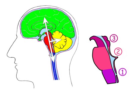
The brainstem assures the link between the spinal cord (blue) and the cortex (green). It derives from the three main vesicles: the myelencephalon, which gives rise to the medulla oblongata (1), the metencephalon, which gives rise to the pons (2) and the mesencephalon (3). It is also a compulsory pathway between the cortex and the cerebellum (yellow).
NB. for a general schematic of the auditory pathway, see the page “Overview”
From the cochlea to the brainstem
P. Gil-Loyzaga
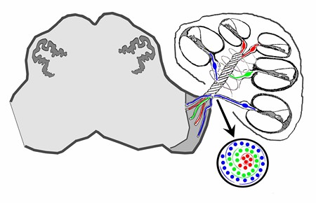
The auditory pathway begins in the spiral ganglion of the cochlea, where the axons of the spiral ganglion neurons form the auditory nerve. The latter, located within the modiolus, is formed by successive layers: central fibres (red) from the apex of the cochlea are progressively surrounded by more basal fibres (blue). In this way, the cochlear tonotopy is maintained.
NB. for a general schematic of the auditory pathway, see the page “Overview”
Les noyaux cochléaires
The cochlear nuclei
The auditory nerve penetrates through the skull and forms a first relay station at the level of the cochlear nuclei.
Nerve fibers entering the cochlear nuclei are distributed in an organised, tonotopic, manner: those that come from the apex of the cochlea (low frequencies) remain at the surface of the cochlear nuclei, whereas those of the base (high frequencies) penetrate deeper into the nuclei. This distribution allows each frequency to activate neurones located in a band that is sensitive to stimulation from a specific frequency known as isofrequency laminae.
Each fiber divides into a “V” shape with two branches: one ventral ascending (for the ventral nucleus) and another dorsal-descending (for the posterior and dorsal cochlear nuclei).
P. Gil-Loyzaga et F. Valderrama
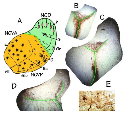
In this diagram, you can see the tonotopic ‘V’
- DCN = Dorsal cochlear nucleus
- AVCN = anterior ventral cochlear nucleus
- PVCN = posterior ventral cochlear nucleus
Types de neurones: Neural types:
- E = Spherical bushy
- Glo = Globular cells
- Es = Stellate cells
- O = Octopus cells
- Gr = Granular cells
- G = Giant cells
- PF = fusiform and pyramidal cells
Function
The cochlear nuclei decode intensity, analyse various temporal parameters such as duration (long or short sounds) and the start and end of a sound stimulus, and maintain and transmit the frequency analysis carried out previously at the level of the cochlea.
The function of each type of neuron is well defined. They have the essential role of completing the frequency and intensity analyses started at the cochlea.
- Globular and spherical neurons have a primary response similar to that of the spiral ganglion neurons: they respond continuously throughout a stimulus
- Octopus cells fire at the beginning of the stimulus (‘onset response’)
- Fusiform and pyramidal cells of the DCN fire at the beginning of the stimulus (‘on-off’ response).
The cochlear nuclei have numerous internal circuits with excitatory or inhibitory connections between neurons, which contribute to the analysis of the auditory signal and its distribution towards the higher centres of the auditory system, starting with the the next relay centre: the superior olivary complex.
Projections from the cochlear nuclei
Certain neurons in the cochlear nuclei project towards other nuclei. These contralateral projections form the acoustic stria.
P. Gil-Loyzaga
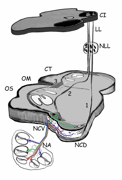
Acoustic Stria
1 – Dorsal stria, or ‘stria of von Monakow’, formed mainly by the nerve fibers of the pyramidal and multipolar cells in the DCN. After crossing over, these fibers project up through the lateral lemniscus (LL) to synapse in the inferior colliculus (IC)
2 – Intermediate acoustic stria or ‘stria of Held’, formed by the axons of the octopus cells in the VPCN which cross the midline and project to the contralateral LL.
3 –Ventral acoustic stria, or trapeziod body. The most complex of the stria, formed by the axons of most neurons in the AVCN (spherical, globular and multipolar), which send their axons bilaterally to the neurons in the superior olivary complex (SOC). The fibers of the olivary neurons project via the LL and synapse primarily with neurons in the IC.