Cochlea
Authors: Mireille Lavigne-Rebillard, Marc Lenoir, Rémy Pujol
Contributors: Sam Irving
In humans fetuses, the organ of Corti begins its differentiation at 9 weeks of gestation (wg). The following set of Nomarski and SEM pictures illustrates the main stages of its development. A timing correspondence between humans and different mammals is given in table I (bottom of the page).
Stage 1 : First signs of differentiation (9-10 weeks of gestation (wg))

M. Lavigne-Rebillard
")
M. Lavigne-Rebillard
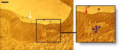
The coiling of the cochlear spiral has occurred (left). However, the sensory epithelium is not yet differentiated (right) – only a ridge and some filaments of the nascent tectorial membrane (arrows) mark its location.
scale bars: 1 mm (left) and 25 µm (right)
![]()
In a transverse Nomarski section of the otocyst, a thick undifferentiated epithelium is seen covered by a thin tectorial membrane (white arrow). The spiral ganglion (red arrow) is more advanced in its development and a track of nerve fibres (blue arrow) can be followed up to the otocyst.
scale bar: 75 µm
(M. Lavigne-Rebillard)
Stage 2 : First signs of hair cell differentiation (11-12 wg)

SEM surface of the newly differentiating hair cells.
The nascent round-shaped bundles of stereocilia from both IHCs (i) and OHCs (o) are seen jutting out above the surrounding microvilli that cover the surface of the support cells.
scale bar: 10 µm
M. Lavigne-Rebillard
Nomarski section from cochlea of an 11 wg fetus. The white arrow points to the tectorial membrane.
scale bar : 20 µm
Insert: swollen nerve endings (blue arrows in inset) are concentrated below the newly differentiating hair cells. A red arrow points to the IHC.
Stage 3 : Organ of Corti just prior to the onset of function (14-15 wg)

M. Lavigne-Rebillard
SEM surface of a 14 wg human fetus cochlea.
Stereocilia of both IHCs (i) and OHCs (o) have grown and the tufts have lost the round-shaped pattern.
scale bar : 10 µm

M. Lavigne-Rebillard
Nomarski section from a 14 wg human fetus.
IHC (red arrow) and OHCs (blue arrows) are clearly distinguishable on both sides of the differentiating pillar cells.
scale bar : 20 µm
Similar developmental stage in a 2 postnatal day (pnd) old mouse.
Note the inner sulcus filled with the thick Kölliker’s organ (asterisk) which plays a major role in forming the tectorial membrane by secretion (white arrow).
scale bar : 20 µm

M. Lenoir
Stage 4 : Structural stage corresponding to the onset of cochlear function (18-20 wg)

R. Pujol
In all mammals studied, this stage corresponds to the onset of cochlear function (see table I).
At this stage (here a pnd 8 mouse), the tunnel of Corti is open, Nuel’s spaces are forming beside the OHCs (blue arrows), and Kölliker’s organ (asterisk) is regressing, freeing the inner spiral sulcus and the tectorial membrane.
In this section, the IHC is not fully visible.
scale bar : 15 µm
Supernumerary hair cells
M. Lavigne-Rebillard

M. Lavigne-Rebillard
Des Supernumerary hair cells are frequently and temporarily encountered during the development of the human organ of Corti, between stages 3 and 4 ( ref c4). As shown here (14 wg, left; 20 wg, right) 2 rows of IHCs (red arrows) and 4 or 5 rows of OHCs (blue arrows) are often seen, even in the basal turn.
scale bar: 20 µm
Stage 5 : End of morphological differentiation of the organ of Corti (30 weeks’ gestation in the human)

M. Lenoir
SEM picture (rat organ of Corti).
At the end of its development (30 weeks of gestation) the human organ of Corti looks like this one (taken from a one month-old rat).
Note an almost complete disappearance of microvilli on the surface of the support cells, especially those of the pillar cells.
scale bar : 15 µm
Mature organ of Corti from a guinea-pig (Nomarski).
The main differences between stages 4 and 5 concern the final stages of differentiation of the OHCs and surrounding structures.
scale bar : 10 µm
")
R. Pujol
Table I: timing of maturation of the organ of Corti in different species
This table gives comparative timings of stages 2 (onset of hair cell morphological differentiation), stage 4 (onset of cochlear potentials), and stage 5 (end of maturation) in different mammals (ref b21).
wg = weeks of gestation (humans) ; ed = embryonic days ; pnd = postnatal days
| species | stage 2 | stage 4 | stage 5 |
| Human Rat, Mouse Cat Guinea-pig Gerbil |
10 wg 16-17 ed ? (ed) 34 ed ? (ed) |
18 wg 8-10 pnd 3 pnd 54 ed 12 pnd |
30 wg 16-20 pnd 20 pnd 6 pnd 20 pnd |
Table II: comparative development of the cochlea and the auditory brain in both humans and rats
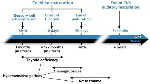
This table recalls stages 2,4, and 5 (see above) of cochlear maturation and highlights the late development of the auditory brain. Actually, to mature properly, the brain requires the cochlea to be fully mature and functional.
The hypersensitive periods to three major deleterious factors of cochlear development are indicated. During these periods the cochlea is much more susceptible than in adulthood.