Inner Ear
Authors: Rémy Pujol
Contributors: Sam Irving
The inner ear corresponds to 2 distinct sensory organs: the balance organ or vestibule, and the hearing organ or cochlea, which have a similar embryological origin, the otic vesicle. Both organs also share morphological and physiological properties, such as hair cells and the transduction mechanism.
Vestibule and cochlea: two sensory organs of the inner ear
Two sensory organs are located in the inner ear. The vestibule is the organ of equilibrium and the cochlea the organ of hearing. They share a common embryonic origin (otic vesicle), plus different morphological or physiological properties such as endolymph, hair cells and mechano-transduction.
The schematic drawing below represents the osseous (top left) and membranous (seen by transparency in the main drawing) labyrinths.
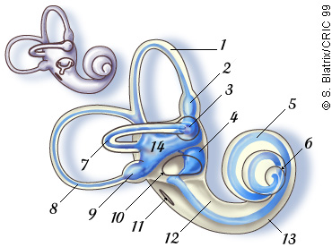 1. Anterior semicircular canal
2. Ampulla (superior canal)
3. Ampulla (lateral canal)
4. Sacculus
5. Cochlear duct
6. Helicotrema
7. Lateral (horizontal) canal
8. Posterior canal
9. Ampulla (posterior canal)
10. Oval window
11. Round window
12. Vestibular duct (scala vestibuli)
13. Tympanic duct (scala tympani)
14. Utricule
14. Utricule
Sur ce schéma, on visualise l’étroite relation anatomique entre les organes vestibulaires et la cochlée. On voit, en particulier, la continuité endolymphatique (bleu) qui permet de comprendre que toute pathologie affectant l’endolymphe ait des répercussions sur les deux fonctions sensorielles. Ex. hydrops, certains types de maladie de Ménière.
In situ schematic drawing of the human inner ear
The bone has been removed to visualise the vestibule (1), the VIIIth nerve (2) , and the basal portion of the cochlear duct (3), housing the organ of Corti. The rest of the cochlea (4) is covered by the bony capsule. The VIIIth nerve is formed by the vestibular and cochlear nerves which merge before entering the brain.


Human cochlea (5 months fetus)
The bony capsule has been dissected out, showing the 2 1/2 coils of the membranous labyrinth (35 mm in length). The oval (blue arrow) and round (yellow arrow) windows are indicated..
Picture: Mireille Lavigne-Rebillard
Scale bar: 0,5 cm