Tous droits réservés © NeurOreille (loi sur la propriété intellectuelle 85-660 du 3 juillet 1985). Ce produit ne peut être copié ou utilisé dans un but lucratif.
 Français
 English
 Español
 Português
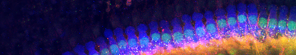
Both types of hair cells are innervated by specific afferent and efferent systems forming a loop to and from the brainstem.
Innervation of inner (1) and outer hair cells (2)
 et externes (2)")
|
The radial afferents (blue) and the lateral efferents (pink) innervate the inner hair cells; the spiral afferents (green) and the medial efferents (red) innervate the outer hair cells. |
IHC innervation

The IHC is synaptically connected to all type I spiral ganglion neurons (refs. a1, c5) forming the radial afferent system (blue) going to the cochlear nuclei (CN). The lateral efferent system (pink) arising from small neurons in the ipsilateral lateral superior olivary complex (LSO) brings a feedback control to the IHC/type I afferent synapse.
OHC innervation

The OHC synapses with a few (at least in basal and mid-portions of the cochlea) small endings from type II spiral ganglion neurons (ref. c1), forming the spiral afferent system (green). In turn, large neurons of the medial efferent system (red), from both sides of the medial superior olivary complex (MSO), form axo-somatic synapses with the OHC.
Schematic representation of the hair cells afferent innervation

|
Hair cells afferent innervation is made by peripheral fibers of the spiral ganglion neurons (SGNs) Type I (blue) SGNs (95% of the ganglion neurons) have a single ending radially connected to IHCs (as a mean, 10 SGNs/IHC). Type II (green) small, unmyelinated neurons spiral basally after entering the organ of Corti and branch to connect about a tenth OHCs, generally in the same row. Scheme from Liberman |
The spiral (cochlear) ganglion
The spiral ganglion is formed from the primitive otocyst. It differentiates very early, before the organ of Corti. In man, it is composed of 30 to 35,000 bipolar neurons of two main types as described above. Both types have central axons (which form the auditory nerve) delivering messages to the cochlear nuclei.
See
specific pages for details on SGN anatomy and physiology.
Facebook Twitter Google+