Spiral Ganglion: anatomy
Conception : Rémy Pujol
The spiral ganglion is formed from the primitive otocyst. It differentiates very early, before the organ of Corti. In man, it is composed of 30 to 35,000 bipolar spiral ganglion neurons (SGNs) of two main types. Large and slightly myelinated type I neurons (accounting for more than 90%) are connected to inner hair cells; small and unmyelinated type II neurons are connected to outer hair cells. Both types have central axons delivering messages to the cochlear nuclei ( see also).
The spiral (cochlear) ganglion

Echelle : 10 µm

Echelle : 5 µm
Three type I SGNs (blue arrows) and one type II SGN (green arrow) are seen, together with sections of myelinated fibers (axons from neighboring neurons).
Scale bar: 10 µm
Two type I SGNs and one type II SGN (arrow)
Scale bar: 5 µm
Type-I SGN slightly myelisated by its glial satellite cell
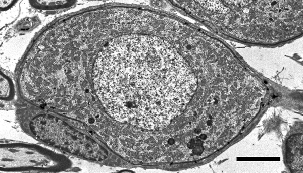
This type I SGN (guinea-pig cochlea) is ensheathed by processes from a satellite glial cell, which form a thin myelin sheath (note the difference with the axons myelin sheaths below). Type I SGN have a the clear cytoplasm full of mitochondria, indicating the high metabolic status of the cell. On the right is the axon hillock.
Scale bar: 3 µm
Type-II SGN and its glial cell
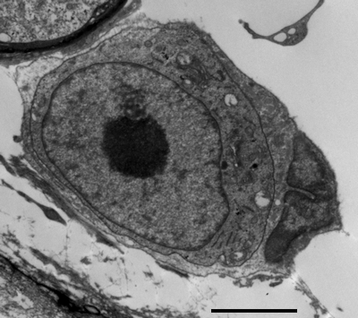
Type II SGN (guinea-pig cochlea). Smaller than type I neurons, type II SGN shows a denser cytoplasm, but less mitochondria. It is surrounded by a glial cell, which do not form myelin.
Scale: 1 µm.
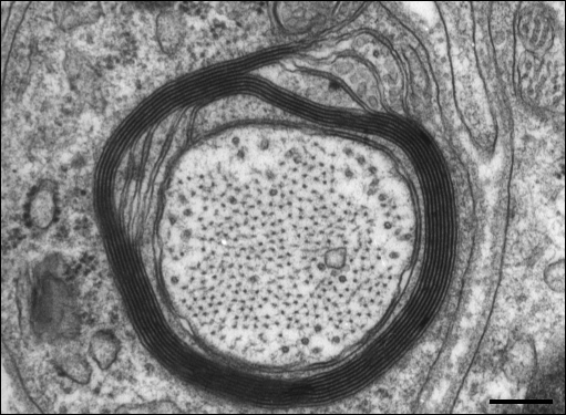
Pre-ganglionic myelinated fibers (peripheral axons from type I SGN)
Cross-section of a myelinated osseous spiral lamina fibre (peripheral axon from a type I neuron). Within the axon, 3 mitochondria are seen within numerous microtubules and filaments. The myelin sheath is formed of about 30 layers (see electronic zoom).
Scale bar: 150 nm
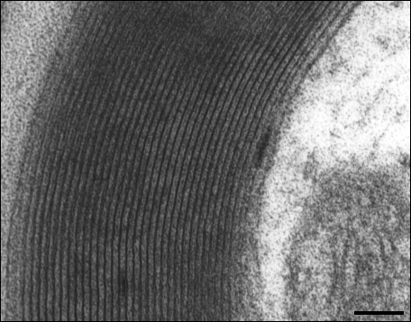
35 turns of the myelin sheath are visible. The axon cytoplasm is seen to the right. Scale bar: 70 nm
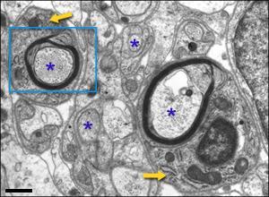
During development, intra-ganglionic auditory fibres (asterisks), are ensheathed by a glial cell process. Two of these glial cells (yellow arrows) have already formed a myelin sheath of several layers (see electronic zoom).
Scale bar: 0,5 µm
An auditory axon, with microtubules and neurofilaments, is being ensheathed by glial cell processes which are forming about 7 to 8 layers of myelin (compare with the adult sheath).Scale bar: 100 nm