Tous droits réservés © NeurOreille (loi sur la propriété intellectuelle 85-660 du 3 juillet 1985). Ce produit ne peut être copié ou utilisé dans un but lucratif.
 Français
 English
 Español
 Português
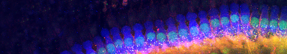
This synaptic organisation is characteristic of the cochlear region that encodes very low frequencies: this is more extensive in humans (representing almost all of the apical turn). Everything occurs as though these cochlear OHCs remained ‘true’ sensory cells, perhaps sending messages to the brain to reinforce the perception of low frequencies.
Base / Apex differences
The base-apex difference could potentially be explained by the examination of what happens during synaptogenesis.
Everything occurs as though synaptic development beneath the OHCs adapted to the physiological properties of these cells: The apical OHCs maintain a ‘classic’ sensory function (with innervation similar to that of the IHCs), whereas the basal OHCs, having become electromotile, are now equipped with a system to regulate this new function.

R. Pujol |

|
Synaptic poles of an OHC (o) at the apex of the cochlea: start of the fourth turn (above) and extreme apex (below) in the guinea pig.
In contrast to the basal turn, the synaptic pole ot the OHC is essentially surrounded by afferent dendritic boutons.
The vesiculated efferent terminals (red arrows) are very infrequent and the postsynaptic differentiation incomplete (blue arrow).
d = Deiters' cell.
scale bar: 1 µm and electronic zoom on the right

R. Pujol |

|
N.B. This synaptic organisation is characteristic of the cochlear region that encodes very low frequencies: this is more extensive in humans (representing almost all of the apical turn). Everything occurs as though these cochlear OHCs remained ‘true’ sensory cells, perhaps sending messages to the brain to reinforce the perception of low frequencies ?
OHC synaptogenesis
The base / apex difference can be explained by the development of the OHCs: see the diagram of synaptogenesis below.
Everything occurs as though synaptic development beneath the OHCs adapted to the physiological properties of these cells: The apical OHCs maintain a ‘classic’ sensory function (with innervation similar to that of the IHCs), whereas the basal OHCs, having become electromotile, are now equipped with a system to regulate this new function.
Thus, a convergence of messages from both IHCs and OHCs may account for a quite good sensitivity for low frequencies, despite the undevelopment of the active mechanism.

|
Stage 1 – The base of the immature OHC synapses only with the type II spiral ganglion neuron afferents (in green) and the radial fibres (type I, in blue), the latter with numerous synaptic bodies. |

|
Stage 2 – At the moment of the functional beginnings of the cochlea, the first efferent synapses are visible (in red), whereas the number of radial afferent fibres (in blue) decreases. Compare this stage with the synaptic organisation of an OHC at the apex of an adult cochlea (see diagram below). |

|
Stage 3 – Once the cell is mature, the classic OHC synaptic organisation can be observed: a large efferent terminal (in red) and a small type-II afferent terminal (in green) are present. This organisation is characteristic of the OHCs in the region which encodes for high- and mid- frequencies (i.e. in the majority of the cochlea). |
Facebook Twitter Google+