Tous droits réservés © NeurOreille (loi sur la propriété intellectuelle 85-660 du 3 juillet 1985). Ce produit ne peut être copié ou utilisé dans un but lucratif.
 Français
 English
 Español
 Português
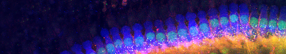
Since the large myelinated type I Spiral Ganglion Neuron (SGN) make up the vast majority (95%) of SGNs, they are the only type described in details here. Their neural activity can be recorded using a sharp micro-electrode inserted into the auditory nerve. In an adult cochlea, the type I SGN peripheral fiber form a single bouton synapsing with one inner hair cell (IHC), and recordings reflects a precisely located activity.
Both, spontaneous activity (in absence of sound) and evoked activity (to sound stimulation) can be recorded.
Spontaneous activity of auditory nerve fibers
In absence of sound stimulation, a SGN initiates spontaneous spikes.
The spontaneous rate (SR) of discharge varies a lot from one SGN to another and depends on certain molecular characteristics. Three categories of SGN have been described depending on their SR:
- low-SR (less than 0.5 spike/sec), about 15% of the SGNs, all forming synapses on the modiolar side of the IHC.
- high-SR (over 18 spikes/sec), about 60% of the SGNs, forming synapses on the tunnel pillar side of the IHC.
- in-between, an intermediate class called medium-SR represents about 25% of the SGN population.
In this video, three SGNs coming from the same cochlear region show 3 different SRs: in green, a low-SR fiber, in red a high-SR fiber, in blue a medium-SR fiber. See below for the respective function of these 3 pools of fibers in coding the sound intensity.
Evoked responses of auditory nerve fibers
Type I spiral ganglion neuron have a relatively narrow auditory receptive field (frequency bandwidth around 1 or 2 octaves) and a limited dynamic range to sound level (around 20-30 dB). As a consequence, to encode sound information over the whole range of the auditory field (10 octaves in frequency, 120 dB in intensity in humans), a great number of SGNs is needed (remember the ratio of 10 SGNs / 1 IHC !).
The auditory nerve fibers show a large variety of responses to frequency and intensity. Fibers specialized for low- and high-frequency sounds innervate respectively the apex and the base of the cochlea. Fibers with a low-threshold have a high spontaneous activity and, conversely, fibers with a high-threshold have low spontaneous activity.
Coding the sound intensity
For a given frequency (in this example, 4 kHz), the 3 types of fibers, coming from the same location in the cochlea, and characterized by their SR, combine their response to code precisely the range of intensity.

- The high-SR (red) responds at threshold (8 dB-SPL here) and significantly increases its response, while its latency decreases, up to 30-40 dB.
- The medium-SR (blue) starts responding around 30 dB and significantly increases its response, while its latency decreases, up to 50-60 dB.
- The low-SR (green) starts responding around 50 dB and significantly increases its response, while its latency is decreasing, up to 80 dB.
The rate/level curves on the right illustrates this mode of cooperation: for the given frequency, when the high-SR fiber response reaches a plateau, the relay is taken by the response of the medium-SR fiber; similarly, when the medium-SR response reaches a plateau, it is relayed by the low-SR fiber response. Overall, a precise coding of the intensity up to 80 dB is sent to the brain.
Tuning curve of auditory nerve fibers
To determine the characteristic frequency (CF) of a fiber, a pure tone can be used as a probe. By varying the frequency and the intensity of the probe, it is possible to assess the receptive field of the neuron.When the probe enters into the receptive field, the neuron increases it discharge rate (red color). Outside the receptive field, the activity of the neuron goes down to the spontaneous rate (blue color).
This is how is built a tuning curve from a neuron recorded in a Mongolian gerbil, with a characteristic frequency around 4.5 kHz and a threshold around 2 dB SPL. Note that the receptive field narrows as the intensity lowers, to become punctiform at threshold.
Temporal response of auditory nerve fibers
In response to a tone-burst presented at the CF, the temporal response of high-CF auditory nerve fibers (CF > 2 kHz, fibers that innervate the basal part of the cochlea) is characterized by a good synchronization at the beginning of the stimulation, an adaptation and a steady-state activity. Low-CF auditory nerve fibers (CF < 2 kHz, fibers innervating the apical part of the cochlea) phase-lock with the waveform of the tone.
This is an exemple of the temporal coding for a fiber from the base of the cochlea (purple, CF = 20 kHz), and a fiber from the apex (yellow, CF = 0.4 kHz). See more about phase-locking and intensity coding at IHC Physiology
Facebook Twitter Google+