OHCs: coupling
Authors: Rémy Pujol
Contributors: Sam Irving
OHC electro-motility is responsible of the active mechanism. However, a tight coupling between OHC and the surrounding structures is needed to knock-on the created strenght to the IHC and the sensory transduction.
At high frequency levels, both sides of the OHCs are coupled. Their base fits in a Deiters’cell cup, which in turn is firmly linked to the basilar membrane. On the other end, the tallest OHC stereocilia are anchored in the tectorial membrane.
This coupling is decreasing from base to apex, where an active mechanism is not really needed for low frequencies.
Mechanical coupling between OHCs and surrounding structures
 Schematic representation
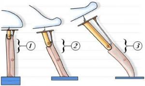
Stages 1 to 3 represent the coupling to their surrounding structures of OHCs in the bat (very tight), in the basal portion of a guinea pig cochlea, and in the apex of the guinea pig cochlea (very loose). In an individual cochlea, the strength of this mechanical coupling follows a decreasing gradient from base to apex – the higher the frequency, the stronger the coupling. This gradient accounts for the efficacy of the reverse transduction process (active mechanism). See also the variation of OHC length (the ” xylophone“).
Legend. In the scheme above as well as in the enlargements below: OHC in yellow, basilar membrane in dark blue, Deiters’ cell in pink with microtubules in brown, tectorial membrane in light blue.

Bat cochlea
In this cochlea, specialised for coding very high frequencies, a very short OHC (yellow), about 10 µm in length, is half-ensheathed in the Deiters’ cell cup; a dense fascicle of microtubules (brown) in the Deiters’ cell links the cup with a thick basilar membrane (dark blue).
- a) The tallest stereocilia are deeply embedded in a massive tectorial membrane (light blue) parallel to the BM.
- b) In the space between the membranes of both the OHC and the Deiters’ cell dense material and septa are seen.
Together, these features account for a very firm coupling and the active mechanism can feedback energy even above 100 kHz.

Basal turn of a guinea-pig (or human) cochlea
At a place coding for frequencies around 10 to 20 kHz, the Deiters’ cup is more like a close-fitting seat: one free side of a longer OHC (around 20 to 30 µm) being occupied by the characteristic huge medial efferent endings.
- a) The tallest OHC stereocilia are not so deeply embedded in the TM.
- b) The extracellular material (and septa) in the Deiters’ cell cup is less obvious.
Compared to the bat, the fascicle of microtubules in the Deiters’ cell remains dense whilst the phalangeal process is longer; both TM and BM are thinner than in the bat

Apical turn of a guinea-pig (or human) cochlea
In an area coding below 1 kHz, an elongated OHC (>70 µm) is now very loosely sited on a Deiters’ cell, which is almost devoid of microtubules and has a very long and thin phalangeal process; both TM and BM are much thinner and they form a much wider angle (no longer parallel).
- a) Even the tallest OHC stereocilia are not embedded with the TM (no imprints in the TM are seen).
- b) The Deiters’ cup is now devoid of dense material.
Coupling between OHC and Deiters’ cell
Transmission electron microcrographs (TEM) shows detailed feature of the coupling.
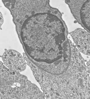
OHC from the upper basal turn of a rat cochlea. The OHC fits in the Deiters’ cell cup.
The OHC innervation is not visible in this section.
This type of junction is enlarged in the following images.

Within the Deiters’ cell (d), a fascicle of microtubules (red arrows) is seen at the junction with the OHC (o).
As shown in the schemes above, the microtubules fascicle runs through the Deiters’ cell, down to its junction with the basilar membrane.
scale bar: 500 nm
")
A higher magnification of this junction between OHC (clear) and Deiters’ cell (dense) shows material and septa (yellow arrows) linking both membranes.
scale bar: 150 nm
Coupling between OHC and tectorial membrane

Scanning electron micrograph (SEM) of the inferior face of the rat tectorial membrane.
Imprints of the tallest OHCs stereocilia are clearly seen.
scale bar: 10 µm

In this transmission electron micrograph, the tallest stereocilium of an OHC is just detached (red arrow) from the tectorial membrane by the fixation procedure.
scale bar: 1 µm