Complexe olivaire supérieur
Conception : Pablo Gil-Loyzaga, Rémy Pujol
L’olive supérieure, ou plutôt le complexe olivaire supérieur (COS), est, sur le plan anatomique et fonctionnel, une des structures les plus complexes de la voie auditive.
Il est composé de plusieurs noyaux, et des zones péri-nucléaires, qui forment un réseau neuronal intense et avec des nombreuses projections ascendantes (vers les noyaux supérieurs de la voie auditive) et descendantes (vers les noyaux cochléaires et la cochlée). Le COS est impliqué dans l’analyse complexe et le filtrage de l’information auditive qui monte vers le cortex cérébral ; il l’est aussi dans l’élaboration de reflexes importants pour la survie de l’individu et la protection du système auditif.
Enfin, le COS joue un rôle important dans la localisation de la source sonore, mais comme il partage cette fonction avec d’autres structures (le colliculus inférieur notamment), cela fera l’objet d’une page spécifique.
Organisation du complexe olivaire supérieur (COS)
Le COS est un centre majeur d’intégration et de régulation de l’information auditive ascendante et descendante.
- Il reçoit des informations du récepteur auditif par les noyaux cochléaires,
- Il analyse ces informations dans leurs propres circuits neuronaux, même pour localiser le point d’origine d’un son dans l’espace entourant l’individu,
- et il renvoie des informations hautement transformés à de nombreuses structures neurales.
Le COS est constitué par un grand nombre de neurones disposés dans les noyaux et les champs péri-nucléaires, qui sont symétriques de part et d’autre de la ligne médiane.
P. Gil-Loyzaga
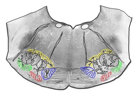
Coupe frontale du pont (zone inférieure) au niveau du COS.
Sur cette vraie coupe anatomique (cerveau de rat), l’olive latérale (en S), l’olive médiane et la strie du corps trapézoïde ont été surlignés.
Les noyaux périphériques sont indiqués en couleur : – en vert , le noyau latéral du corps trapézoïde NLCT, – en rouge, le noyau ventral du corps trapézoïde NVCT, – en bleu, le noyau médian du corps trapézoïde NMCT.
Les principaux types de neurones, impliqués dans le résumé fonctionnel qui va suivre sont aussi positionnés.
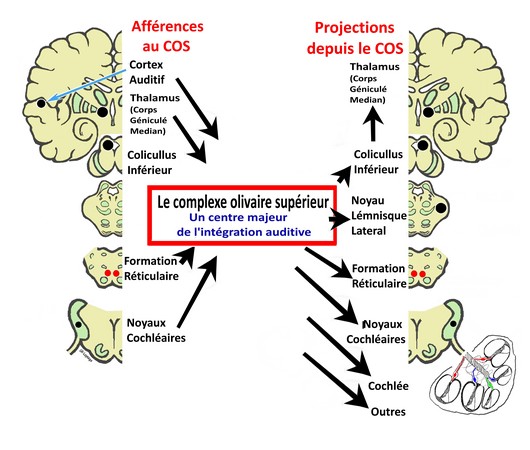
Le COS : afférences et projections
P. Gil-Loyzaga
Le COS reçoit l’information auditive en provenance des noyaux cochléaires et, après l’avoir traitée, la transmet au colliculus inférieur.
L’activité du COS est contrôlée et régulée par :
- les relais supérieurs de la voie auditive : colliculus inférieur, corps genouillé médian (thalamus) et cortex auditif ;
- les noyaux cochléaires ;
- la formation réticulée,
Le COS envoi aussi des projections :
- à la formation réticulée, pour impliquer les informations auditives dans les réactions végétatives, les systèmes d’alerte, l’éveil cortical, etc.,
- aux noyaux cochléaires, effectuant un retro-contrôle de l’activité de ces noyaux,
- à la cochlée (systèmes efférents latéral et médian),
- au noyaux du nerf facial (réflexe stapédien) et du nerf trijumeau (réflexe du marteau),
- au noyau du nerf oculomoteur latéral pour participer au repérage visuel de la source sonore.
Tonotopie au niveau du COS
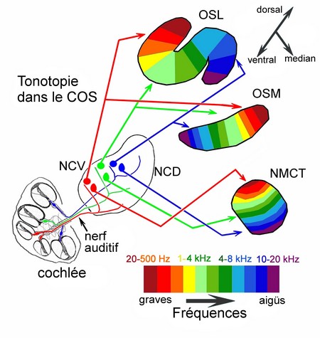
P. Gil-Loyzaga (d’après Guinan and Irvine)
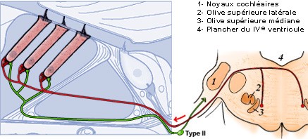
Les neurones du ganglion spiral (cochlée) ont des projections tonotopiques dichotomiques sur les noyaux cochléaires (NCV= Noyau cochléaire ventral; NCD = noyau cochléaire dorsal).
Les neurones sphériques du NCV envoient des projections à l’olive supérieure latérale (OSL) et médiane (OSM) du COS, alors que les neurones globulaires du NCV font des contacts avec les neurones globulaires du NMCT (noyau médian du corps trapézoïde).
Les 3 noyaux du COS montrent une distribution tonotopique très typique : avec les fréquences graves dans les aires dorsales ou latérales et les fréquences aiguës dans les aires plus ventrales ou internes.
Circuits protecteurs et modulateurs des activités périphériques
Les neurones du COS envoient des fibres qui descendent (faisceaux efférents) vers les récepteurs auditifs et établissent des circuits de type réflexe permettant une modulation ou une protection (notamment contre le bruit excessif). Ces neurones efférents, sont eux-mêmes sous contrôle des voies descendantes des centres auditifs supérieurs.
Circuit modulateur neurones auditifs primaires (système efférent latéral)
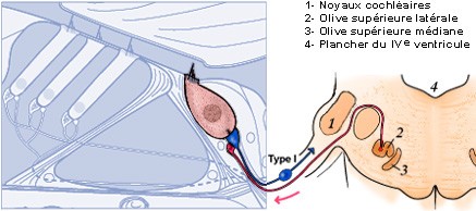
S. Blatrix / R. Pujol
Les CCIs font synapse avec tous les neurones de type I du ganglion spiral, formant le système afférent radial (= nerf auditif : bleu) qui relie la cochlée aux noyaux cochléaires. C’est par là que partent tous les messages auditifs vers le cerveau. Le système efférent latéral (rose), issu de petits neurones de l’olive supérieure homolatérale (2), effectue un rétro-contrôle sur la synapse CCI/fibre afférente.
Ce circuit efférent intervient notamment en libérant de la dopamine pour protéger cette synapse contre un accident excitotoxique (ischémie ou trauma).
Circuit modulateur de l’activité des CCE (système efférent médian)
S. Blatrix / R. Pujol
Les CCE font synapse avec des terminaisons dendritiques de petite taille des neurones ganglionnaires de type-II, formant le système afférent spiral (vert) ; rien n’est connu à propos du rôle de ce système : peut être informe-t-il le système nerveux de l’état de contraction (électro-motilité) des CCE ?
En retour, les CCE sont directement innervées par les grosses terminaisons axoniques (rouge) de neurones situés bilatéralement dans le complexe olivaire supérieur médian (MSO) : c’est le système efférent médian (dont le rôle est de modérer l’électromotilité (mécanisme actif) des CCE.
N.B. Pour mieux visualiser les fibres, les cellules de Deiters n’ont pas été dessinées.
Circuits de protection au niveau de l’oreille moyenne
Si l’intensité d’un son (ou bruit) est trop élevée, il peut y avoir des dommages, parfois irréversibles, des récepteurs auditifs, avec surdité et/ou acouphènes.
Pour éviter cette possibilité le système auditif utilise des circuits réflexes de protection.
Ces circuits impliquent : les noyaux du COS et d’autres noyaux moteurs du tronc cérébral qui activent la contraction des muscles de l’oreille moyenne.
Il exite deux réflexes complémentaires, agissant sur la chaine ossiculaire, qui peuvent réduire l’intensité du bruit (jusqu’à l’annuler).
- Le réflexe acoustique, ou réflexe stapédien (le plus important chez l’homme) implique le nerf facial qui déclenche la contraction du muscle de l’étrier. Il est mis en jeu par des sons d’intensité supérieur à 80 dB.
- Le réflexe du muscle du marteau active la contraction de ce muscle après stimulation du nerf trijumeau (V nerf crânien). Ce réflexe agit contre les sons de très forte intensité, bien que chez l’homme semble avoir une importance secondaire.
Fonctionnement
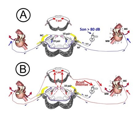
Lorsqu ’un son fort est détecté par la cochlée (> 80 dB) l’information est transmise aux noyaux du tronc cérébral (n. cochléaires et COS). Une boucle réflexe commande la contraction de ces muscles (chez l’homme seul le stapédien se contracte). Ceci entraîne une augmentation de la rigidité de la chaîne tympano-ossiculaire, une limitation des déplacements aux fréquences basses et moyennes (< 2000 Hz) et donc une diminution de l ’énergie transmise à l’oreille interne (par contre, ce réflexe ne protège pas l’oreille aux fréquences élevées).
P. Gil-Loyzaga
A : Circuit du réflexe stapédien
B : Circuit du réflexe du muscle tenseur du tympan
CCI: des cellules ciliées internes, NA: nerf auditif, CN: noyaux cochléaires, NF: nerf facial (VII nerf crânien); NT: nerf trijumeau (V nerf crânien), ME: muscle de l’étrier, MM: muscle du marteau ou tenseur du tympan.
Rôles et limites des réflexes ossiculaires
Ces réflexes ossiculaires réduisent la fonction de transfert entre l’oreille externe et la cochlée ; ils protègent la cochlée contre les surstimulations sonore…mais avec des limites :
- ils sont fatigables : pas de protection lors de bruits de longue durée ;
- ils n’entrent en jeu que pour des fréquences graves (ne dépassant guère 1 kHz);
- ils n’intervienent pas, ou trop tard (latence du reflexe = 30 ms), lors de bruits impulsifs (explosions, armes à feu, pétards, cornes de brume, etc.).
Un autre rôle des réflexes ossiculaires, déclenchés par la vocalisation, est d’atténuer la perception de sa propre voix : ceci est particulièrement important chez les chanteurs.

Vue schématique interne de la cavité de l’oreille moyenne permettant de comprendre comment la chaîne des osselets peut être mobilisée par la commande réflexe des muscles du marteau (9) et de l’étrier (5).
- (1) Marteau
- (2) Ligament du marteau
- (3) Enclume
- (4) Ligament de l’enclume
- (5) Muscle de l’étrier
- (6) Platine de l’étrier
- (7) Tympan
- (8) Trompe d’Eustache
- (9) Muscle du marteau
- (10) Corde du tympan sectionnée