Tous droits réservés © NeurOreille (loi sur la propriété intellectuelle 85-660 du 3 juillet 1985). Ce produit ne peut être copié ou utilisé dans un but lucratif.
Human auditory cortex represents 8% of the surface of the cerebral cortex. Unlike other cerebral areas, there are key structural differences between the auditory cortices of different mammalian species, as well as between apes and humans.
Anatomy of human auditory cortex
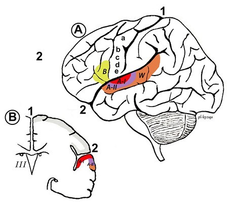
The human auditory cortex can be studied using functional magnetic resonance imaging (fMRI), and is divided into more than a dozen different regions surrounding Heschl's gyrus in the superior part of the temporal lobe. Auditory cortex, narrower in humans than in other mammals, develops from front to back within the Sylvian fissure at the point where it joins Heschl's gyrus.
Primary auditory cortex (AI) is situated in the posterior third of the superior temporal gyrus (also known as Brodmann area 41), next to Wernicke's area (W). AI is the central region of the auditory cortex and receives direct projections from the ascending auditory pathway, particularly the ventral region of the medial geniculate body (MGB) in the thalamus.
Secondary auditory cortex (AII) is located more rostrally in the temporal lobe and contains Brodmann area 42.
Anatomical distribution of the auditory cortex
| A - Lateral view showing the distribution of AI and AII and Wernicke's area (W). Auditory cortex projects to the regions of the frontal lobe involved in motor function for speech (a), the lips (b), jaw (c), tongue (d), larynx (e) and Broca's area (B). B - Frontal view showing AI inside the Sylvian fissure and Heschl's gyrus. 1 - Interhemispherical fissure. 2 - Sylvian fissure |
Structure and circuitry of the auditory cortex: Columnar organisation
The presence of six cell layers in the auditory cortex is common to all mammals, but species differences take the form of the commonality of each cells within each layer. In humans, pyramidal cells (including all types) correspond to 85% of AI. The remaining 15% are multipolar or stellate cells. Inverted stellate cells also exist (Martinotti cells) as well as cells with candelabra-shaped dendritic configurations.
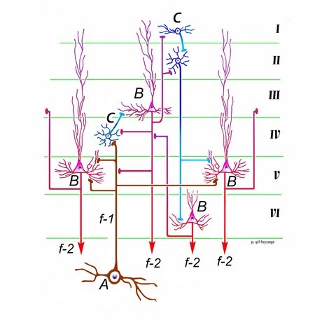
Most ascending fibers originate in the MGB and synapse with the pyramidal cells of layer IV, but this is not always the case. However, these contacts represent only 20% of the excitatory fibers that project to cortical neurons: the other 80% comes from other neurons in the ipsilateral cortex.
Neurons in AI and AII are functionally organized into columns, first described by Lorent de Nó. Cortical columns receive input from both MGBs and are therefore bilateral, working on the principal of summation/suppression. Summation corresponds to similar afferentation from both ears, with a contralateral dominance. Suppression is ipsilaterally dominant.
Cellular organisation and circuitry of the human auditory cortex
| Each neuron of the MGB that projects to the auditory cortex (C) generates a fiber (f-1) that branches horizontally for a few millimeters and contacts numerous pyramidal cells (B) and puncta (C). This system allows the amplification of the auditory signal and improved analysis of its activity. Neurons in layer IV project to the pyramidal neurons of layer III, and from there the information is distributed to the other layers (I, II, IV and V) of the ipsilateral cortex and the contralateral auditory cortex via the corpus callosum. Layer I neurons project to layer II, which in turn connect with layers V and VI. Pyramidal neurons in layers V and VI have efferent axons (f-2) that project to the MGB. Those of layer V also project to the inferior colliculus. All of these neurons also send collateral connections back up to layers III and IV. |
Specificity of the human auditory cortex
While other levels of the auditory pathways are very similar within species, the human neocortex is characterized by the predominence of pyramidal cells (85% of cortical neurons), and some very specific types of cells such as inverted pyramidal cells and candelabrum neurons. Another specificity is the massive interconnection of cortical neurons, which accounts for 80% of excitatory synapses in the neocortex. Only 20% are coming from the medicular genicular body !
 Français
 English
 Español
 Português


Facebook Twitter Google+